
 |
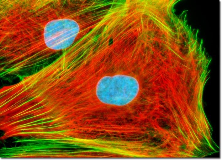
| Image Above: Embryonic rat myoblast dyed with mouse and goat antibodies. (4) |
adenocarcinoma, however, type 2 diabetes being a metabolic consequence is
supported both by experimental and clinical data. In addition, in vivo
and in vitro experiments have shown pancreatic cancer cells can alter muscular
and liver glucose metabolism, as well as cause peripheral insulin resistance
(making natural insulin less powerful against reducing blood glucose levels).
Lastly, the researchers also wanted to take a look at cachexia ( a
symptom of pancreatic cancer [cachexia is known as wasting syndrome; loss of
weight, muscle atrophy, fatigue, etc.; this is a symptom that is common to a
lot of cancers, as well]) and see its relationship to glucose metabolic
expression. From here the researchers were going to verify whether
pancreatic cancer cell conditioned media affected glucose metabolism in mice
myoblasts and to compare the gene expression of mice myoblasts exposed to
pancreatic cancer media versus non- exposed mice myoblasts. To compare gene expression and find which genes were being altered a microarray experiment was set up a platform of 5000 skeletal muscle cDNA. (1)
To run this experiment, many different aspects of
the experiment had to be covered. The first part of the experiment
required setting up conditioned media. For the conditioned media,
researchers used four different human pancreatic cancer cell lines and one
colorectal cancer cell line, in order, for the researchers too see if something
was specific to pancreatic cancer or general to cancer as a whole. For the
analysis of myoblasts being effected by pancreatic cancer, thousands of mice
myoblasts were plated and cultured. Using these myoblasts (myoblasts were
either exposed to a pancreatic/colorectal cancer strain or were in the
non-exposed myoblasts) the researchers ran a series of six experiments: three
with U-13C-Glucose ( a glucose with a tracer on it) and three with
lactate. In these experiments lactate and glucose data were collected using
a colorimetric method on an automatic analyser. Lastly, for the
microarray experiment, mice myoblasts were plated on petri dishes, allowed to
incubate, glucose and lactate production was measured, the genes were
fabricated using PCR, hybridized, and validated using a combination of PCR and
gel-electrophoresis. (1)
Results
Their results were stunning. Researchers
found that in the conditioned myoblasts versus non-conditioned myoblasts,
glucose concentrations declined slightly in all experimental
 conditions.
However, one specific pancreatic cancer conditioned myoblast (BxPC3) line
was statistically significantly different compared to non- conditioned
myoblasts, after 48 hours of incubation. The figure to the right is
showing the different non- conditioned andpancreatic cancer lined myoblasts
measured from 0 to 72 hours for the tracer glucose. This part of the
researcher's experiment verified that pancreatic cancer cells do indeed effect
the glucose metabolism of mice myoblasts. (1)
conditions.
However, one specific pancreatic cancer conditioned myoblast (BxPC3) line
was statistically significantly different compared to non- conditioned
myoblasts, after 48 hours of incubation. The figure to the right is
showing the different non- conditioned andpancreatic cancer lined myoblasts
measured from 0 to 72 hours for the tracer glucose. This part of the
researcher's experiment verified that pancreatic cancer cells do indeed effect
the glucose metabolism of mice myoblasts. (1)
For the microarray portion of the experiment,
researchers found that there is a large number of over- and underexposed genes
in cancer conditioned myoblasts versus non-conditioned myoblasts. Some of
these genes also can be linked to being involved in the modification of the
glucose metabolism. (1)
Overall, the researchers found that media
conditioned with pancreatic cancer cells increased lactate production and
induced proteolysis, the process of breaking down proteins into smaller
polypeptides or completely into amino acids. These processes likely were
caused by pancreatic cancer cells altering the expression of a large number of
genes-- that were involved in protein biosynthesis and glucose metabolism. (1)
Analysis
1) Given these results do we buy them? Are
the statistics there?
Yes we do. For starters, on the statistical
side of things they have a huge paragraph in the article about how these
results will be statistically analyzed using a one way ANOVA and Bonferroni's
test for comparisons. Solely that is not enough though, the researchers
actually use these statistical tests in each figure provided in every figure
caption. In addition to these statistical tests, the researchers had a
PCR and gel electrophoresis set up to give them the ability to validate the
results from the microarray experiments. (1)
Using these methods of analysis the data is credible from a statistical
stand point.
2) Would the experimenters be able to reproduce the
experiment? Why do the researchers choose myoblasts, as their cell of choice?
In my opinion, yes they would. There has been
a large amount of discussion on diabetes and its relation to pancreatic cancer
and this study attempts to verify those claims with a test of the direct
effects of glucose metabolism on cells. I will admit though, while this
experiment is very interesting and a great break through it would have been
nice to have seen the experiment ran on some sort of organism's beta cells (a
type of pancreatic cells) or even hepatocytes (liver cells). In general,
the only problem ( or abnormality) in running this experiment is the use of
muscle cells because it seems odd not to use a type of pancreatic cells, like
beta cells, as these would make a lot more sense given the large focus of other
research on the early onset of diabetes and its link to pancreatic
cancer. Another point that needs to be made is even in the discussion
section of this journal article the researchers bring up that gene
alterations they observed may also occur in other target cells like insulin
secreting cells. (1) So why did they not test beta cells?
3) Taking genes to a new level.
Now that these researchers have identified a lot of
genes that may be the cause of altered glucose metabolism in myoblasts there
should be more tests done to find conclusive evidence of which specific genes
are the ones causing the modified glucose response (the researchers in this
experiment just found a bunch of genes that could possibly identify the affect
on glucose metabolism, not specific genes). Once specific genes are
identified, researchers could use this information to formulate some sort of
gene therapy to correct the over- and under expression of these genes, which
has the potential to be a very interesting, breakthrough approach to correcting
some of the symptoms of pancreatic cancer like cachexia. As the
researchers mentioned in the discussion, cachexia can be induced by glucose
intolerance and atrophy.
4) A link to class.
After reading this experiment, thinking about gene
expression, and what we have discussed in class thus far, I wonder
if maybe pancreatic adenocarcinoma involves an onocogene, and a symptom of
the proto-oncogene being mutated to an oncogene leads to a change in the
genome, much like the viral oncogene Peyton Rous discovered. Oncogene's
usually involve increased cell division or inhibition of cell death (2), but, maybe this oncogene somehow causes
glucose metabolism altering genes to be over- and under expressed.
Another and more likely alternative could be the oncogene expressing an
increase in glucose receptors, if this happens this could somehow lead to a
high concentration of glucose in the blood, which is known as hyperglycemia or
diabetes. This makes even more sense, as doctors have already proven there
is some link between type 2 diabetes and pancreatic cancer. This would
be a truly amazing breakthrough, however, this is a very big leap of faith and
it would take a lot more research to prove or even delve into this subject
matter if this was a possible mechanism of pancreatic adenocarcinoma.
5) Future experiments
An example future experiment that could be done
(besides more tests to clarify specific genes affecting the gene expression)
using a very similar technique could be replacing the mice myoblasts with a
sample of human myoblasts or a type of pancreatic cells from humans.
However, I know nothing of the feasibility of getting either of these
cells, as I assume some sort of biopsy would have to be performed, and unless
the human was dead getting a sample of pancreatic cells from a living human
would be quite invasive and probably not worth the risk (one exception could be
if they were having surgery on the pancreas, say a pancreatomy, then it could
be a much more worthwhile/easier process, as the surgeons would already be
inside the person's torso removing part or all of the pancreas).
Final thoughts
Overall, this experiment brings great insight to
the effect of pancreatic cancer on glucose metabolism in cells. It was
also great to see the different statistical tests the researchers used to
validate their results. One other point to make about the validity of this
article is this data is slightly out of date, as this experiment was done in
2004. I will keep researching to hopefully find some newer data on this
experiment. However, thus far I have been unsuccessful in finding similar
data. I even ran a search on the Pancreatic expression database [(3),(6)]
[a database set up by the Barts Cancer Institute located in London] to try and
find some similar data and could not find any data that was similar to the
experiment these researchers performed. In addition to searching the database,
I found two papers [(5), (7)] that were somewhat similar in talking
about gene expression, but their focus was on the differences in gene
expression of pancreatic cancerous tissue opposed to healthy pancreatic tissue,
not about gene expression and glucose metabolism. Not finding any other
research like this journal article, leads me not to discredit their findings,
but I would prefer a few more studies, before we full heartedly believe in this
data and use it for bigger implications, like gene therapy.
Thanks for reading,
-- Quincy
Here is the article for those who want to read
further:
Citations
1) Basso, D. "Altered Glucose Metabolism and
Proteolysis in Pancreatic Cancer Cell Conditioned Myoblasts: Searching for a
Gene Expression Pattern with a Microarray Analysis of 5000 Skeletal Muscle
Genes." Altered Glucose Metabolism and Proteolysis in Pancreatic
Cancer Cell Conditioned Myoblasts: Searching for a Gene Expression Pattern with
a Microarray Analysis of 5000 Skeletal Muscle Genes. GUT: An International
Journal of Gastroenterology and Hepatology, 03 Feb. 2004. Web. 30 Apr.
2012.<http://gut.bmj.com/content/53/8/1159.full>.
2) Chial, H. (2008) Proto-oncogenes
to oncogenes to cancer. Nature Education 1(1)
3) Claude Chelala, Stephan A Hahn, Hannah J
Whiteman, Sayka Barry, Deepak Hariharan, Tomasz P Radon, Nicholas R Lemoine and
Tatjana Crnogorac-Jurcevic, Pancreatic Expression database: a generic model for
the organization, integration and mining of complex cancer datasets. BMC Genomics
2007, 8:439 [PDF]
4) Davidson, Michael W. "Embryonic Rat
Thoracic Aorta Medial Layer Myoblast Cells (A-10)." Fluorescence
Digital Image Gallery. Florida State University, 14 Oct. 2004. Web. 29 Apr.
2012. http://micro.magnet.fsu.edu/primer/techniques/fluorescence/gallery/cells/a10/a10cellsexlarge18.html
5) Grützmann R, et al. Gene
expression profiling of microdissected pancreatic ductal carcinomas using
high-density DNA microarrays. Neoplasia. 2004;6:611–622.
6) Rosalind J. Cutts, Emanuela Gadaleta, Stephan A.
Hahn, Tatjana Crnogorac-Jurcevic, Nicholas R. Lemoine and Claude Chelala, The
Pancreatic Expression database: 2011 update. Nucleic Acids Research (Database
Issue). 2010, 1-6. [PDF]
7) Shen, Jianjun, Maris D. Person, Jijiang Zhu,
James Abbruzzese, and Donghui Li. "Protein Expression Profiles in
Pancreatic Adenocarcinoma Compared with Normal Pancreatic Tissue and Tissue
Affected by Pancreatitis as Detected by Two- Dimensional Gel Electrophoresis
and Mass Spectrometry." CANCER RESEARCH 64 (2004):
9018-026. Web. 30 Apr. 2012.
<http://www.biochemie.uni-greifswald.de/vorlesungen/Seminar/wahlfach_tumorbiologie/ProteinExpressionProfilesinTumors.pdf>.